Hearty Insights: Recent Advances in the Cardiac Field
Coming off the heels of this year’s Basic Cardiovascular Sciences Scientific Sessions (BCVS 2024), the following publication review focuses on an array of cardiac research, ranging from human ...
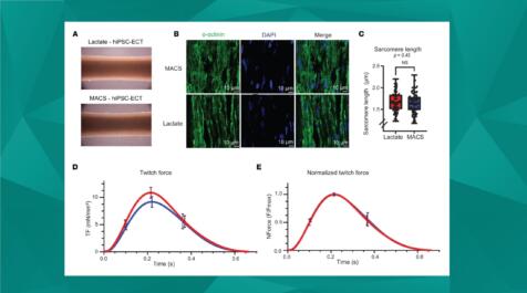
Renewed Interests: Harnessing Engineered Constructs to Explore Novel Strategies in Regenerative Medicine
Tissue engineering is at the forefront of regenerative medicine, offering immense promise for restoring or enhancing the function of impaired tissues and organs. While tissue constructs remain costly ...
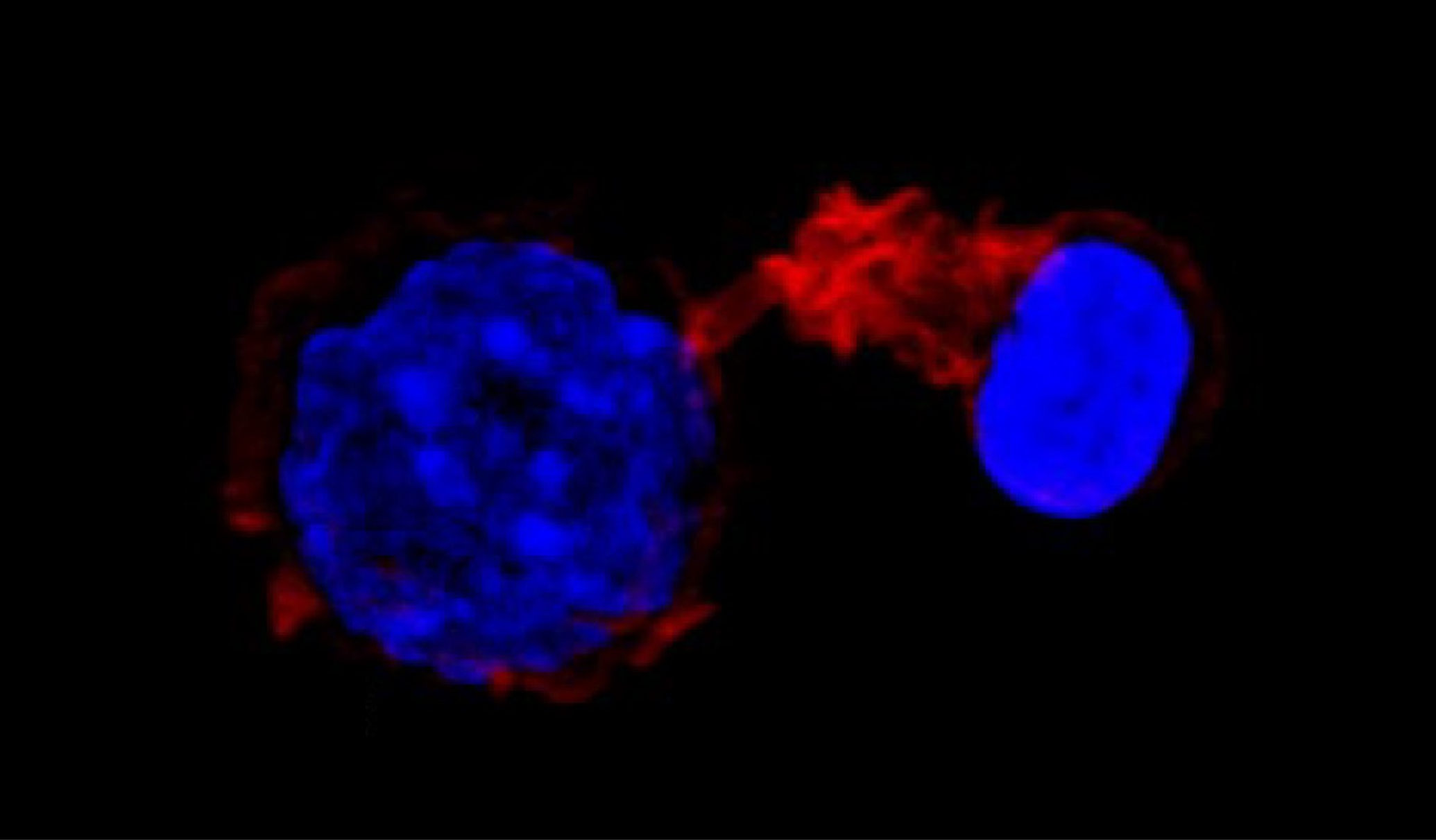
Influence of external forces on actin-dependent T cell protrusions during immune synapse formation
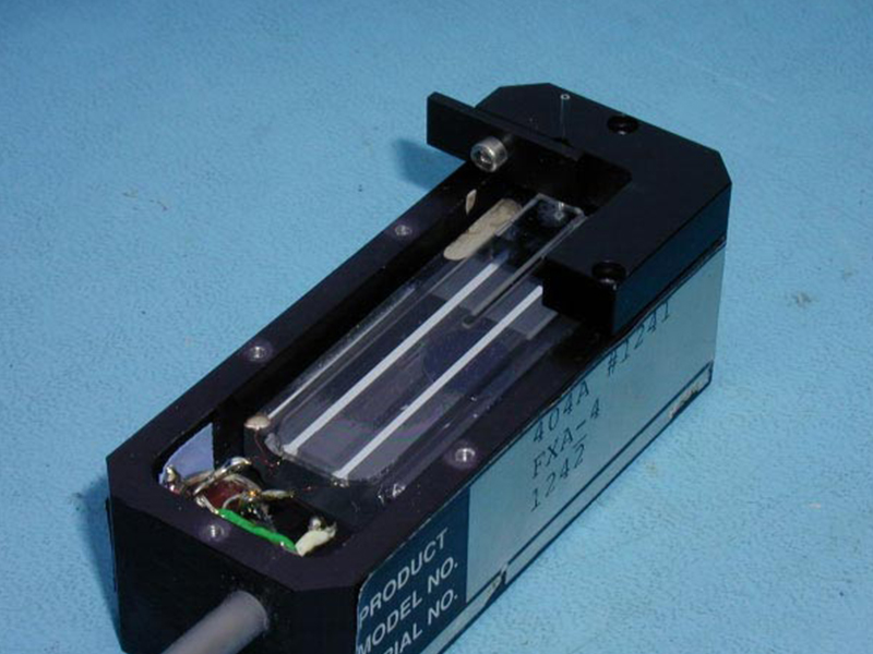
T-cell activation is necessary for producing an adaptive immune response. For T-cell activation to occur, an antigen-presenting cell (APC) must make direct contact with it. This contact region has been shown to control many T-cell functions, and it is from this contact point that protrusions have been observed. To further characterize these protrusions, this study investigated the effect of various APC-mimicking forces on protrusion dynamics. To mimic APC-induced T-cell activation, a micropipette force probe attached to an activating microbead was used. To evaluate micropipette flexibility, the bending stiffness was measured against a microindenter calibrated with Aurora’s 406A force probe. T-cell activation was induced once the microbead would make contact with the cell at a desired compressive or pulling force. To determine if protrusion was affected by external forces produced by APCs during activation, protrusion lengths were monitored following the application of various forces. It was found that the higher the compressive force, the shorter the protrusion was. Conversely, the larger the absolute pulling force was, the longer the protrusion grew. It was also observed longer protrusions were smaller in diameter, at all forces. To determine whether protrusion dynamics were affected by Arp2/3, a protein complex regulating actin polymerization, T-cells were treated with an Arp2/3-specific inhibitor. Not only were protrusion maximum lengths reduced following treatment, but they began growth later in time and at slower speeds. Finally, confocal imaging was used to assess F actin localization within protrusions. The results showed that the walls were rich with F actin whereas the center was depleted. A similar analysis of T cells and model APCs showed the same F actin localization. This study suggests that protrusion growth is set by an intracellular constant time and that protrusion dynamics are influenced by external forces. In addition to this, actin assembly within protrusions is facilitated by the Arp2/3 complex.

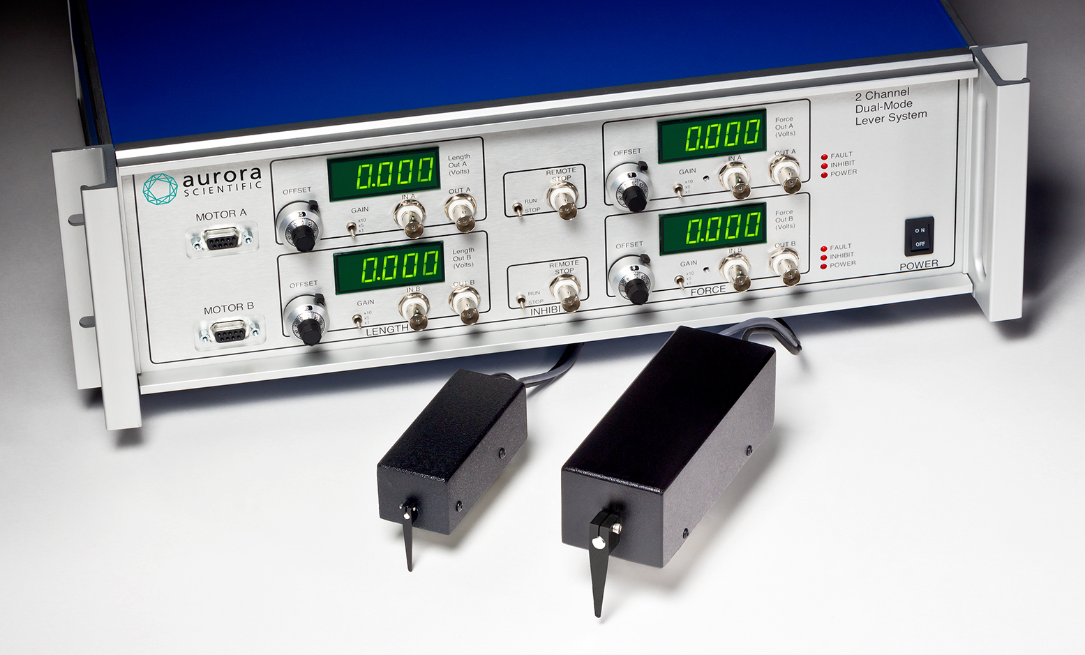
Tips and Tricks for Troubleshooting the 300C Dual-Mode Muscle Levers
Here we will cover [...]
Actuator Design and Testing for Dynamic Applications
For researchers int[...]
Tips and Tricks for Measuring Muscle Function in-vitro
Aurora Scientific’s[...]
Sometimes two is better than one…
Muscle levers and f[...]
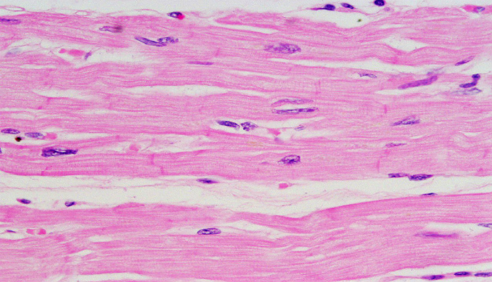
Advancements in Cardiac Tissue Engineering
As our collective u[...]
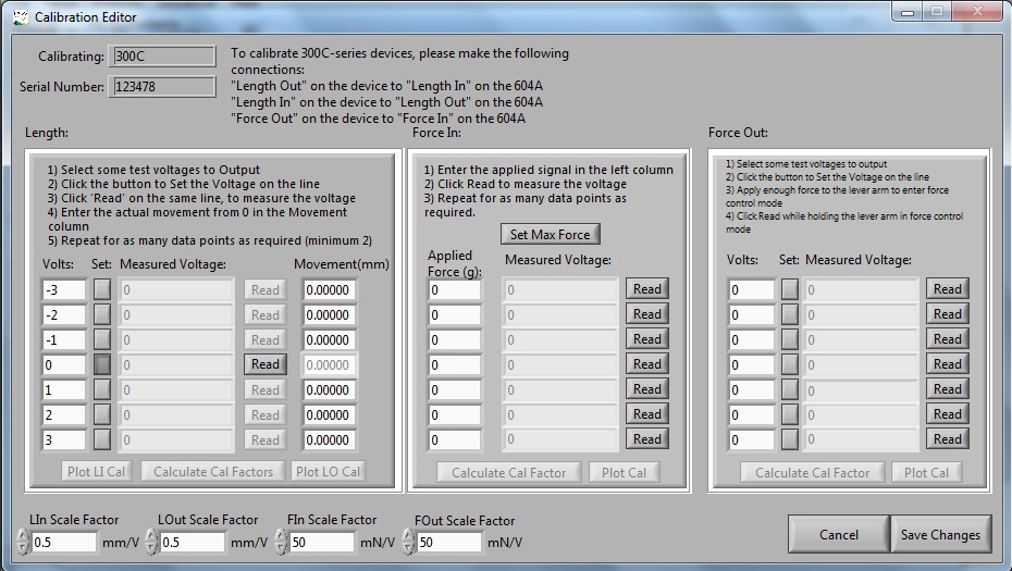
How to Calibrate Your Dual-Mode Lever System Using DMC
This blog is intende[...]

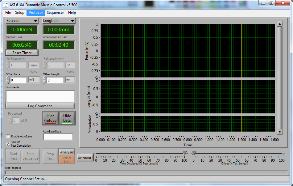
How to Write Basic Protocols in DMC – Isometric
Looking to character[...]
Performing a Tube Repair of an ASI 400A Force Transducer
The procedure for r[...]